

Overview
Cartilage is a smooth and durable tissue that coats the ends of each bone. This type of cartilage is called articular cartilage. It is necessary for healthy joints.
Damage and breakdown of the articular cartilage in the knee happens for many different reasons. Genetics, trauma, age, weight, and activity are some of the potential causes of cartilage injury. When there is breakdown of the cartilage the knee can become inflamed and painful, this is called arthritis.
When arthritis worsens and non-operative treatment fails to reduce symptoms, partial knee replacement or total knee replacement is often the only answer to relieve pain and restore function.
However, there are certain circumstances when we are able to restore and preserve articular cartilage.
In general this is possible when the articular degeneration is localized and isolated to one area of the joint.
Below we will discuss a few operative techniques that address this difficult problem.
Cartilage Restoration Techniques

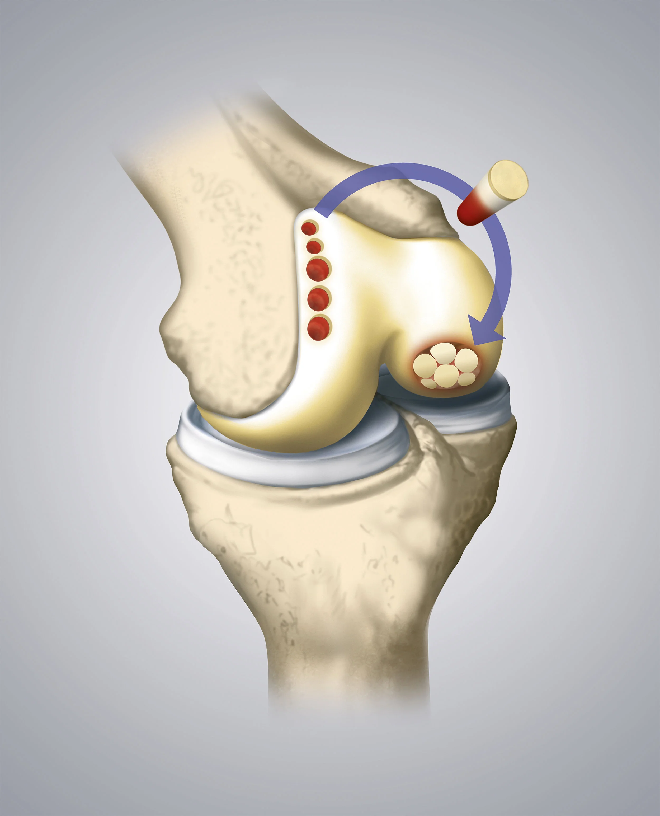
Microfracture
Microfracture is a technique used to help encourage your body’s own cells to migrate to the area of cartilage loss. Small holes are created in the bone at the site that lacks cartilage. Stem cells travel up through these holes from the bone marrow and fill the defect. These stem cells will then transform into cartilage cells. This can work for smaller defects.
Autologous Chondrocyte Implantation (ACI)
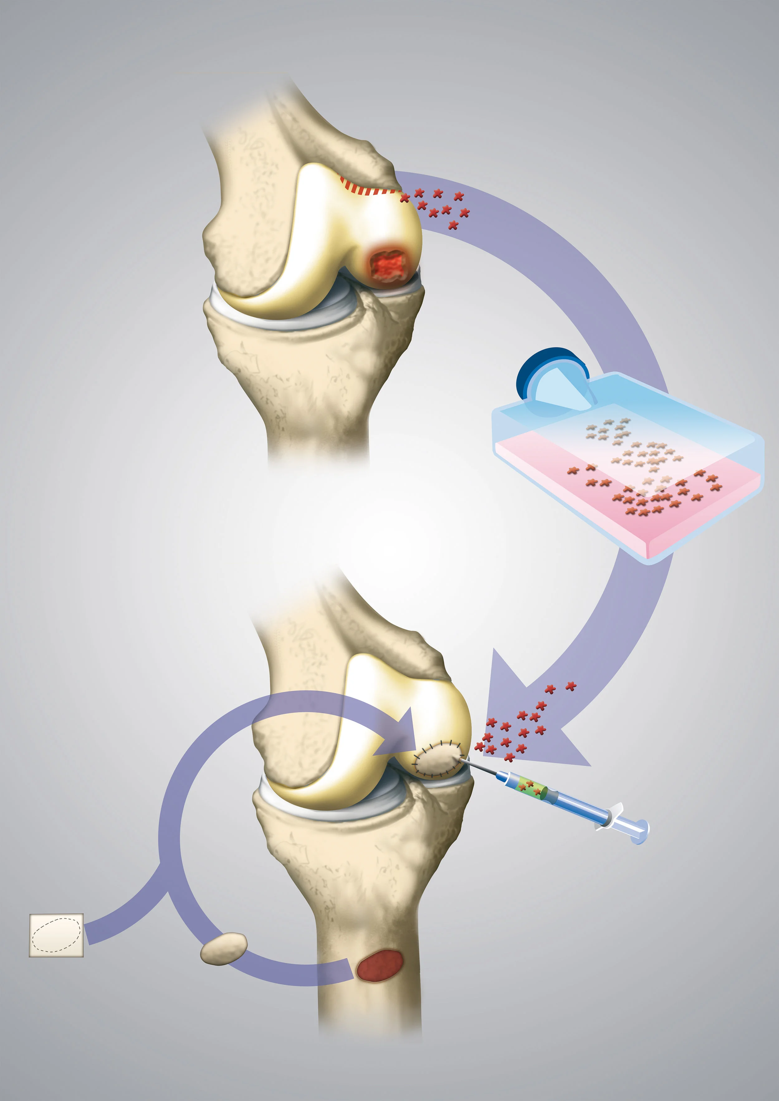
Autologous Chondrocyte Implantation requires two surgeries but has the advantage of using the patient’s own cells.
During the first surgery, healthy cartilage cells, called chondrocytes, are harvested from the knee and sent to a laboratory. The specialized laboratory will then culture these cells in a special medium. The chondrocytes will then proliferate, growing in number. The cells are then placed into a special membrane and shipped back to the surgeon. This process takes several weeks.
During the second procedure, the membrane with the large population of chondrocyte cells are implanted into the defect.
Autograft Transplantation
In osteochondral autograft transplantation (often referred to as an OATS procedure) healthy cartilage is harvested and moved from one area of the knee (donor site) to the area with the arthritic defect.
The donor cartilage comes from a location in the knee that is “less important.”
Sometimes a single plug can fill the defect or occasionally multiple plugs are placed in a mosaic pattern. This procedure is also referred to as “mosaicplasty.”
This procedure is best done with smaller arthritic defects because we are unable to remove too much from the donor site.
Allograft Transplantation

In contrast to autograft transplantation, which is taking cartilage from another location in your own body, allograft transplantation involves implanting cartilage from someone who has elected to donate their cartilage upon death.
The graft is sterilized and tested to reduce the risk of disease transmission.
Allograft transplantation is best for situations with larger areas of cartilage loss.
Summary
Cartilage restoration techniques described above work best in younger patients with isolated areas of arthritis. With advanced arthritis, that affects multiple ares of the knee, the above techniques are not viable options, and knee replacement is a better solution.